Electron Microscopy Captures Individual Nanoparticles: Imagine being able to see the tiniest building blocks of our world—nanoparticles—as clearly as you see the pages of a book. Thanks to a recent breakthrough in electron microscopy, scientists can now capture and study individual nanoparticles with a level of detail and precision never before possible. This remarkable advancement is not just a technical feat; it’s opening doors to new discoveries in medicine, electronics, energy, and beyond.

In this article, we’ll explore what this breakthrough means, how it works, and why it matters—for both curious young minds and seasoned professionals. We’ll break down the science, offer real-world examples, and provide practical advice for those eager to use or understand this technology.
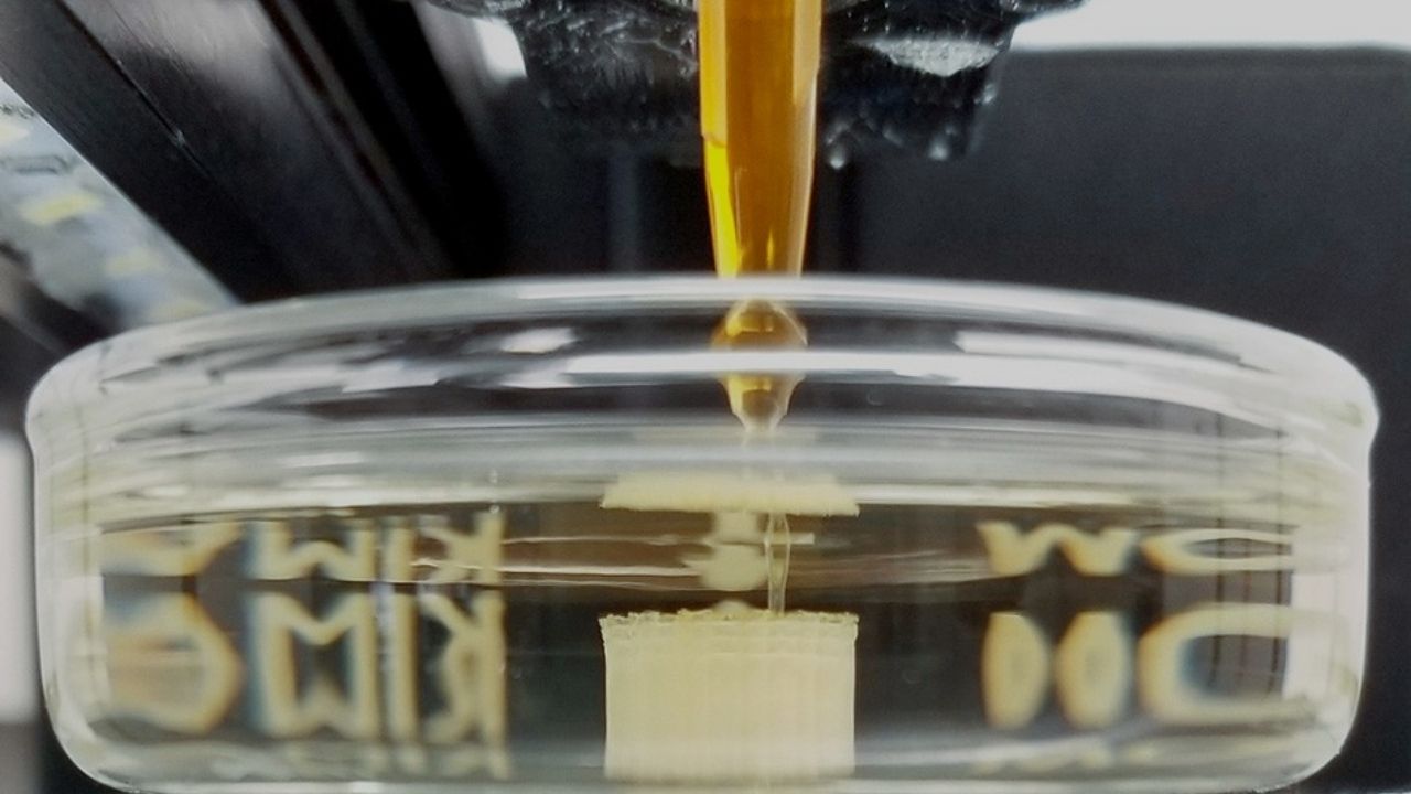
Electron Microscopy Captures Individual Nanoparticles
| Feature/Topic | Details & Data | Professional/Career Impact |
|---|---|---|
| Breakthrough | Real-time 3D imaging of individual nanoparticles using advanced electron microscopy | Enables atomic-level material design, crucial for R&D in nanotechnology, materials science, and pharmaceuticals |
| Precision | Captures atomic changes at 0.02 nanometers—6x smaller than a hydrogen atom | Allows monitoring of chemical reactions and dynamic processes at atomic scale |
| Technology | Combines AI with electron microscopy (Graphene Liquid Cell TEM, Brownian Tomography) | AI skills and microscopy expertise in high demand for research and industry |
| Applications | Pharmaceuticals, electronics, energy, quantum computing, catalysis | Expands opportunities in nanomaterials, device engineering, and drug discovery |
| Career Pathways | Materials scientist, nanotechnologist, data scientist, electron microscopist | Interdisciplinary skills in physics, chemistry, engineering, and AI |
The breakthrough in electron microscopy that allows scientists to capture individual nanoparticles with unprecedented precision is transforming how we understand and engineer the materials that shape our world. By combining advanced imaging, AI, and innovative sample preparation, researchers can now watch atoms move and react in real time and in three dimensions—a feat once thought impossible.
This leap forward is already impacting industries from medicine to electronics, and it’s opening exciting new career paths for those skilled in science, engineering, and data analysis. Whether you’re a student, a professional, or simply fascinated by the invisible world, this technology offers a glimpse into the future of innovation.
What Is Electron Microscopy and Why Is This Breakthrough Important?
Electron microscopy is a powerful imaging technique that uses electrons instead of light to see objects at the atomic and molecular level. Traditional optical microscopes can’t resolve features smaller than about 200 nanometers, but electron microscopes can visualize structures as small as a few atoms.

Nanoparticles are materials so tiny that they’re measured in billionths of a meter (nanometers). They’re used in everything from medicines and solar panels to computer chips and batteries. But until now, scientists could only see averaged or static images of these particles, not how they changed or moved in real time—especially in liquids, where most chemical reactions happen.
This new breakthrough lets researchers:
- Watch individual nanoparticles move and change in three dimensions.
- See how atoms rearrange during chemical reactions.
- Study materials in their natural, liquid environments—not just in a vacuum.
Why does this matter? Because understanding how nanoparticles behave at the atomic level helps us design better drugs, stronger materials, and more efficient electronics.
How Does the Breakthrough Work?
Let’s break down the science into simple steps:
1. Advanced Electron Microscopy Techniques
Scientists use a special type of electron microscope called Graphene Liquid Cell Transmission Electron Microscopy (TEM). This device sandwiches nanoparticles between sheets of graphene (a super-thin, strong material) and fills the space with liquid. This setup allows nanoparticles to move freely, just like they would in real life.

2. Brownian Tomography
Nanoparticles in liquid jiggle around randomly—a phenomenon called Brownian motion. By capturing thousands of images from different angles as the particles move, scientists can reconstruct a 3D movie of their atomic structure over time.
3. Artificial Intelligence (AI)
AI algorithms process the massive amounts of data from the microscope. They filter out noise, sharpen the images, and help reconstruct the 3D structure of each nanoparticle at every moment. This makes it possible to see atomic-level changes in real time, even when the particles are moving or reacting.
4. Real-World Example: Platinum Nanoparticles
Using this technique, researchers recently watched platinum nanoparticles as they underwent a chemical reaction. They could see exactly when and where atoms left the surface, rearranged, or reattached—a level of detail that was impossible before.
Why Is This a Big Deal? Real-World Applications
This breakthrough isn’t just for scientists in white coats. It has real-world impacts that touch our daily lives:
Medicine
- Drug Discovery: Many new medicines rely on nanoparticles to deliver drugs to the right cells. Understanding how these particles behave in the body can make treatments safer and more effective.
- Vaccine Development: Visualizing how nanoparticles interact with viruses or immune cells can speed up vaccine research.
Electronics and Computing
- Faster Chips: Computer chips are made from materials structured at the nanoscale. Seeing how atoms move lets engineers design faster, smaller, and more energy-efficient processors.
- Quantum Computing: Atomic-level imaging is crucial for developing quantum bits (qubits), the building blocks of future computers.
Energy and Environment
- Better Batteries: Watching how atoms move in battery materials helps scientists design batteries that last longer and charge faster.
- Clean Energy: Nanoparticles are used in solar panels and fuel cells. Understanding their behavior can make renewable energy more efficient.
Industrial Catalysts
- Chemical Manufacturing: About 90% of industrial products rely on catalysts—materials that speed up chemical reactions. Most catalysts are nanoparticles. Seeing them in action helps companies make cleaner, more efficient processes.
Step-by-Step Guide: How Scientists Capture Nanoparticles in Action
1. Prepare the Sample
- Nanoparticles are placed in a tiny container made from graphene sheets.
- The container is filled with liquid to mimic real-world conditions.
2. Insert into Electron Microscope
- The sample goes into a powerful electron microscope that can see individual atoms.
3. Capture Images
- As nanoparticles move and react, the microscope takes thousands of images from different angles.
4. AI Data Processing
- Advanced AI algorithms clean up the images, removing noise and sharpening details.
5. 3D Reconstruction
- The data is combined to create a 3D movie showing atomic changes in real time.
6. Analyze and Apply
- Scientists study the movies to learn how nanoparticles behave, then use this knowledge to design better materials and products.
Quantum Sensors Now Deliver Unmatched Precision For Medical And Aerospace Breakthroughs
Microsoft Declares the Beginning of the Logical Qubit Era in Quantum Computing
Ultralight 3D-Printed Antennas Cut Weight By 94 Percent With Breakthrough Multi-Material Design
FAQs About Electron Microscopy Captures Individual Nanoparticles
What is a nanoparticle?
A nanoparticle is a tiny piece of matter, usually between 1 and 100 nanometers in size. For comparison, a human hair is about 80,000 nanometers wide!
Why can’t we use regular microscopes to see nanoparticles?
Ordinary microscopes use light, which can’t reveal objects smaller than about 200 nanometers. Electron microscopes use electrons, which have much shorter wavelengths, allowing them to see individual atoms.
What is Graphene Liquid Cell TEM?
It’s a special electron microscopy technique where nanoparticles are observed in a liquid environment, sandwiched between sheets of graphene. This allows scientists to see how nanoparticles behave in real life, not just in a vacuum.
How does AI help in electron microscopy?
AI processes huge amounts of data, removes noise, and reconstructs clear images, making it possible to see atomic-level changes in moving nanoparticles.
What industries will benefit most from this breakthrough?
Pharmaceuticals, electronics, energy, and chemical manufacturing are just a few sectors that will benefit from better understanding and designing nanoparticles.
Where can I learn more or see these images?
Visit the Molecular Foundry at Berkeley Lab or check out Nature Materials for the latest research and images.
Practical Advice: How to Get Involved or Apply This Knowledge
For Students and Young Learners:
- Stay curious! Explore science kits and online resources about nanotechnology.
- Watch videos about electron microscopy to see how scientists work.
For University Students and Professionals:
- Consider courses in materials science, physics, chemistry, or data science.
- Learn about AI and machine learning, as these skills are increasingly important in research.
- Look for internships or research opportunities at national labs or universities with advanced microscopy facilities.
For Industry Leaders and Researchers:
Collaborate with national labs or academic centers to access cutting-edge microscopy.
- Invest in AI and data science training for your teams.
- Stay updated with journals like Nature Materials and organizations like the DOE Office of Science.