Tiny Nanoneedle Cancer-Detection Patch: Imagine if diagnosing cancer or Alzheimer’s disease no longer meant enduring painful, invasive procedures. Thanks to a groundbreaking invention by scientists at King’s College London, that future is now within reach. A tiny nanoneedle patch—covered with tens of millions of microscopic needles, each a thousand times thinner than a human hair—offers a painless, minimally invasive way to collect vital molecular information from tissues, potentially replacing traditional biopsies for millions of patients worldwide.
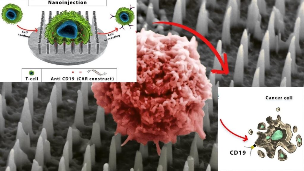
This innovation is more than just a technical leap—it’s a revolution in how we detect and monitor diseases. Unlike standard biopsies, which require cutting or removing tissue and can cause pain, scarring, and complications, the new patch gently extracts molecular “fingerprints” from cells without damaging the tissue. The result? A safer, faster, and far less uncomfortable experience for patients, with the added benefit of being able to sample the same area repeatedly for ongoing monitoring.
Tiny Nanoneedle Cancer-Detection Patch
| Feature | Traditional Biopsy | Nanoneedle Patch |
|---|---|---|
| Pain Level | Painful, invasive | Painless, minimally invasive |
| Tissue Damage | Yes (removes tissue) | No (tissue remains intact) |
| Sampling Frequency | Limited (can’t repeat often) | Unlimited (can sample repeatedly) |
| Speed of Results | Days to weeks | As fast as 20 minutes |
| Disease Monitoring | Difficult | Real-time, continuous |
| Applications | Cancer, Alzheimer’s, etc. | Cancer, Alzheimer’s, brain surgery |
| Manufacturing | Standard medical devices | Semiconductor chip technology |
| Official Reference | — | King’s College London |
The tiny nanoneedle cancer-detection patch developed by King’s College London represents a major leap forward in medical diagnostics. By offering a painless, minimally invasive alternative to traditional biopsies, it has the potential to transform how we detect and monitor diseases like cancer and Alzheimer’s. With the ability to collect rich molecular data without damaging tissue, and the promise of real-time, repeatable testing, this technology could soon become a standard tool in hospitals and clinics worldwide.
For patients, it means less pain and anxiety. For doctors, it means better, faster decisions. And for the future of medicine, it opens up exciting possibilities for personalized, precision healthcare.
How the Nanoneedle Patch Works
Step-by-Step Guide
- Patch Application
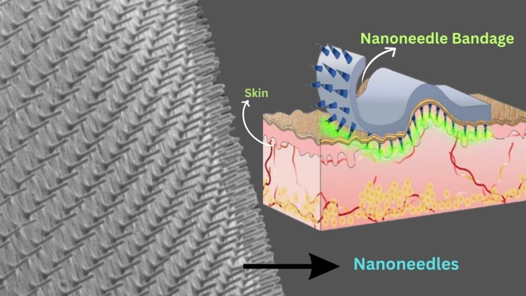
The patch, covered in tens of millions of ultra-fine nanoneedles, is gently placed onto the skin or tissue of interest. The needles are so small that they penetrate the surface without causing pain or damage. - Molecular Extraction
The nanoneedles collect a rich array of molecules—like proteins, lipids, and mRNA—directly from the cells, creating a “molecular fingerprint” of the tissue. This process is called non-destructive sampling because it leaves the tissue unharmed. - Analysis
The collected molecules are analyzed using advanced techniques such as mass spectrometry and artificial intelligence. These tools help identify whether a tumor is present, how it’s responding to treatment, and how the disease is progressing at the cellular level. - Repeatable Testing
Because the tissue isn’t damaged, the same area can be sampled multiple times. This allows doctors to monitor changes over time, providing a much clearer picture of disease progression and treatment effectiveness.
Why This Matters: The Big Picture
For Patients
The nanoneedle patch means less pain, no scarring, and a much lower risk of complications. For children, the elderly, or anyone nervous about medical procedures, this is a game-changer. It also means that sensitive areas—like the brain—can be monitored more safely and frequently.
For Doctors and Scientists
This technology provides multidimensional molecular information from different types of cells within the same tissue, something traditional biopsies simply cannot do. It enables real-time monitoring and more precise decision-making, especially during surgeries. For example, during brain surgery, a patch can be applied to a suspicious area, and results can be obtained in as little as 20 minutes, helping surgeons make faster, more accurate decisions.
For the Future of Medicine
The patch can be mass-produced using the same technology as computer chips, making it easy to integrate into medical devices like bandages, endoscopes, and even contact lenses. This opens up exciting possibilities for personalized medicine, where treatment can be tailored to the unique molecular profile of each patient.
Practical Advice and Real-World Examples
Who Benefits Most?
- Cancer Patients: Especially those with brain tumors, where traditional biopsies are risky and difficult to repeat.
- Alzheimer’s Patients: Early and frequent monitoring could help track disease progression and response to treatment.
- Children and Sensitive Patients: Those who fear needles or have conditions that make invasive procedures dangerous.
What Should You Do?
If you or a loved one is facing a biopsy, ask your doctor about upcoming nanoneedle patch technology. While it’s still in preclinical testing, it’s expected to move to clinical trials soon, and awareness can help you advocate for less invasive options in the future.
Example: Brain Surgery
Imagine a surgeon performing brain surgery. Instead of waiting days for lab results, they apply the nanoneedle patch to a suspicious area. Within 20 minutes, they know exactly what kind of tissue they’re dealing with and can make informed decisions about how much to remove.
Detailed Breakdown: The Science Behind the Patch
Nanoneedles: What Are They?
Nanoneedles are microscopic structures made from porous silicon, each about 1,000 times thinner than a human hair. They’re designed to gently penetrate tissue and extract molecules without causing damage.
How Do They Collect Data?
When the patch is applied, the nanoneedles collect a wide range of molecules from the tissue. These molecules are then analyzed using mass spectrometry and artificial intelligence, which can identify specific biomarkers associated with diseases like cancer and Alzheimer’s.
Why Is This Better Than a Biopsy?
Traditional biopsies remove a piece of tissue, which can be painful and risky, especially in sensitive areas. The nanoneedle patch doesn’t remove tissue, so it’s safer and can be used repeatedly on the same area. This allows for continuous monitoring and more accurate tracking of disease progression.
How Accurate Is It?
In preclinical studies, the patch was able to classify different types of brain tissue (grey matter, white matter, and tumor regions) with high accuracy. Machine learning models using data from the patch matched tissue analysis with up to 85% accuracy in top-ranked correlations, and could predict tumor grade with comparable performance to standard samples.
Biocarbon from Agro‑Waste Serves as Sulfur Host in Next‑Gen Li‑S Batteries
FAQs About Tiny Nanoneedle Cancer-Detection Patch
Q: Is the nanoneedle patch available now?
A: The patch is currently in preclinical testing, but it’s expected to move to clinical trials soon. It’s not yet available for routine use, but the results so far are very promising.
Q: Does it hurt?
A: No, the patch is designed to be painless. The nanoneedles are so small that they don’t cause pain or damage to the tissue.
Q: Can it be used for any type of cancer?
A: The technology has been tested on brain cancer tissue and is expected to be adaptable for other types of cancer and diseases like Alzheimer’s.
Q: How long does it take to get results?
A: In surgical settings, results can be obtained in as little as 20 minutes, much faster than traditional biopsies.
Q: Can the same area be sampled more than once?
A: Yes, because the tissue isn’t damaged, the same area can be sampled repeatedly for ongoing monitoring.
Q: How is the patch made?
A: The patch is manufactured using the same technology as computer chips, making it easy to produce and integrate into medical devices.
